How Vets Diagnose Hip Dysplasia in Dogs
Founder & Veterinary Clinical Advisor at Pets Health Tips · DVM (Doctor of Veterinary Medicine), Member of the American Veterinary Medical Association (AVMA) · 14+ years in companion animal clinical triage, preventative nutrition, and geriatric pet therapy
Hip dysplasia is a silent thief in dogs, it steals mobility slowly, often without obvious signs until it’s severe. Knowing how vets diagnose hip dysplasia in dogs can mean catching it early, before arthritis sets in and your dog’s quality of life drops. This condition affects up to 70% of large breeds like German Shepherds and Labradors, and early diagnosis is the key to managing it.
The Orthopedic Foundation for Animals (OFA) reports that over 100,000 dogs are screened annually in the U.S. alone, with standardized methods to confirm or rule out the condition. Here’s how the process actually works.
how vets diagnose hip dysplasia in dogs
Vets diagnose hip dysplasia through a physical exam and imaging. They check for lameness, gait abnormalities, and joint laxity. X-rays confirm the diagnosis by revealing joint irregularities.
PennHIP and OFA are the two main standardized methods used.
Why Hip Dysplasia Diagnosis Needs a Vet’s Expertise
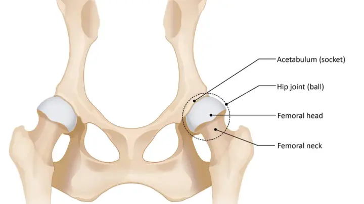
Hip dysplasia isn’t just about a limp or stiffness, it’s a structural problem where the ball-and-socket hip joint doesn’t fit properly. A vet’s trained eye catches subtle signs that owners miss, like a slight "bunny-hop" gait or muscle loss in the hind legs. Without proper diagnosis, dogs can develop painful arthritis, and breeders might unknowingly pass on the condition.
Misdiagnosis is a real risk. Conditions like cruciate ligament injuries or spinal issues can mimic hip dysplasia symptoms. Vets use specific tests, like the Ortolani and Barlow maneuvers, to detect joint laxity that confirms dysplasia.
This precision is why DIY diagnosis or guessing based on symptoms alone often leads to wrong conclusions.
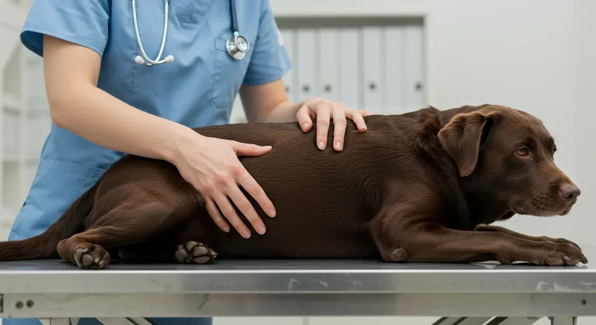
Image source: Bing (Web (fair-use with source credit))
The First Signs That Should Send You to the Vet
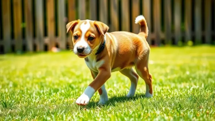
Early signs are easy to overlook. Puppies as young as 4 months may show intermittent lameness or difficulty rising after rest. Older dogs often develop stiffness, especially in cold weather, or reluctance to jump or climb stairs.
These signs don’t always mean hip dysplasia, but they’re red flags that warrant a vet visit.
Some breeds are at higher risk. Large and giant breeds like German Shepherds, Labradors, and Golden Retrievers are genetically predisposed. Even mixed breeds with these lineages can inherit the condition.
If your dog fits this profile and shows any of the above signs, it’s time for a professional evaluation.
Image source: Bing (Web (fair-use with source credit))
How Vets Actually Diagnose Hip Dysplasia (Step by Step)
The Physical Exam: What Your Vet Is Looking For
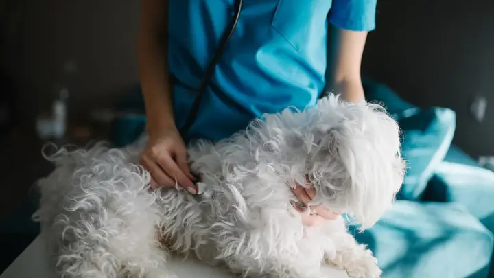
The process starts with a hands-on exam. Your vet will observe your dog’s gait, looking for abnormalities like a swaying walk or "bunny-hopping" when running. They’ll also check for muscle atrophy in the hind legs, which often accompanies chronic hip issues.
Next, the vet performs palpation tests. The Ortolani test involves gently manipulating the hip joint to feel for laxity or a "click" as the femoral head slips in and out of the acetabulum. The Barlow test applies pressure to the joint to assess instability.
Pain or resistance during these tests can indicate dysplasia.
Imaging: X-Rays, PennHIP, and When to Use Each
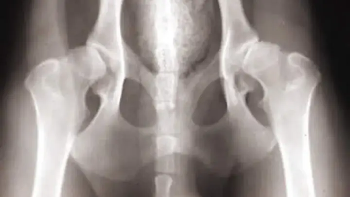
X-rays are the gold standard for confirming hip dysplasia. Standard radiographs, often taken under sedation, show the joint’s structure in detail. Vets look for signs like shallow acetabula, irregular femoral heads, or joint space narrowing.
These images reveal the severity of the condition, which guides treatment decisions.
For early detection, especially in puppies, the PennHIP method is more sensitive. It involves three specific X-ray views, including a distraction view that measures joint laxity. The Orthopedic Foundation for Animals (OFA) method, on the other hand, is typically used for dogs over 2 years old and provides a grading scale from Excellent to Severe.
As of 2026, both methods are widely recognized, but PennHIP is preferred for younger dogs due to its ability to predict future arthritis risk.
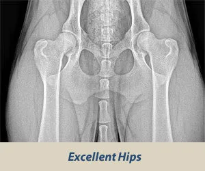
Image source: Bing (Web (fair-use with source credit))
PennHIP vs. OFA: Which Test Is Right for Your Dog?
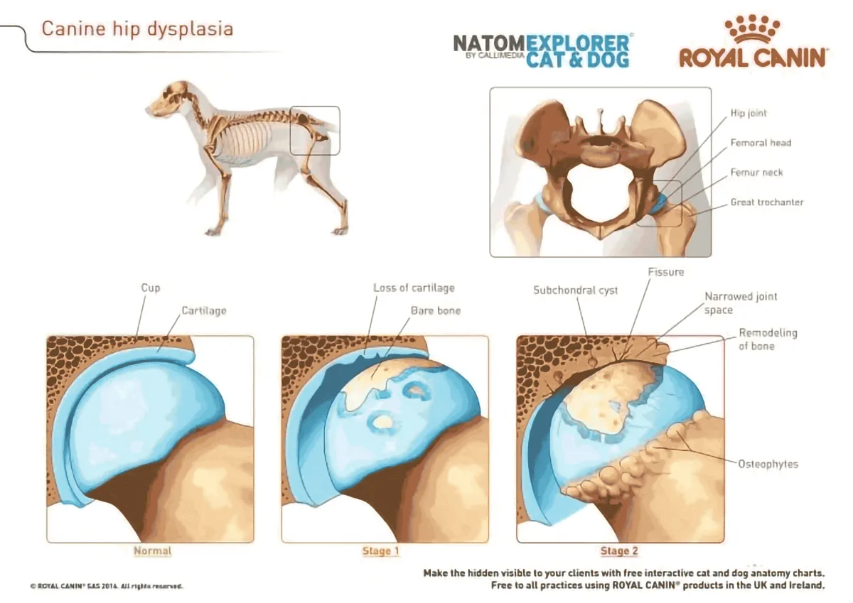
Image source: Bing (Web (fair-use with source credit))
PennHIP and OFA serve different purposes. PennHIP is ideal for puppies as young as 16 weeks and focuses on measuring joint laxity to predict future arthritis. It’s more expensive but provides a distraction index (DI) that quantifies the risk.
OFA, on the other hand, is a pass/fail grading system used for dogs over 2 years old, often for breeding certification.
Here’s a quick comparison:
| Feature | PennHIP | OFA |
|---|---|---|
| Best for | Puppies (16+ weeks), early detection | Adult dogs (2+ years), breeding |
| Cost | $300–$600 | $150–$400 |
| Sedation | Required | Required |
| Turnaround | 1–2 weeks | 2–4 weeks |
| Predictive? | Yes (DI score) | No (grading only) |
PennHIP is the better choice if you want to assess a puppy’s risk or plan early interventions. OFA is the standard for breeders who need official certification. For more details, the OFA’s guidelines provide a full breakdown of their grading system.
What the Results Mean (And What to Do Next)
OFA grades range from Excellent to Severe. A grade of Fair or better is considered passing for breeding purposes. PennHIP provides a distraction index (DI), where a score below 0.3 indicates a low risk of developing arthritis.
If your dog is diagnosed with hip dysplasia, the next steps depend on the severity. Mild cases may be managed with weight control, joint supplements like glucosamine and chondroitin, and physical therapy. Moderate to severe cases might require pain medication, anti-inflammatories, or even surgery such as femoral head ostectomy (FHO) or total hip replacement (THR).
Early intervention is key. Dogs with mild dysplasia can often live comfortably for years with the right care. Regular check-ups and monitoring can help track the progression of the condition and adjust treatment as needed.
Costs, Risks, and How to Prepare Your Dog
The cost of diagnosing hip dysplasia varies. Standard X-rays typically range from $150 to $400, while PennHIP evaluations can cost between $300 and $600. Sedation adds another $50 to $150.
These costs can be higher in specialized clinics or urban areas.
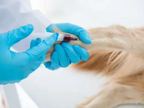
Preparing your dog for the procedure involves a few steps. Your vet may recommend fasting for 12 hours before sedation. A pre-sedation health check, including bloodwork, ensures your dog can safely undergo the procedure.
After the X-rays, your dog may need a quiet space to recover from sedation.
Risks are minimal but worth noting. Sedation carries a small risk of complications, especially in older dogs or those with underlying health issues. Radiation exposure from X-rays is minimal and not a significant concern for a single session.
Always discuss any concerns with your vet before proceeding.

Similar Posts